
Rapidly progressive focal segmental glomerulosclerosis with familial clustering of renal disease.
DOI:
https://doi.org/10.56867/Keywords:
Focal segmental glomerulosclerosis, Rapidly Progressive Glomerulonephritis, Familial clustering, Case reportAbstract
Introduction: Focal segmental glomerulosclerosis (FSGS) is a histopathological pattern of glomerular injury characterized by the presence of sclerosis in a proportion of glomeruli (focal) and partial involvement of the glomerular tuft (segmental). Its etiology is heterogeneous, and it can be classified as primary, genetic, secondary, or of undetermined cause. We present the case of a young adult patient with FSGS, featuring a marked family history of renal disease, resistance to immunosuppressive therapy, and rapid progression to end-stage renal disease requiring renal replacement therapy.
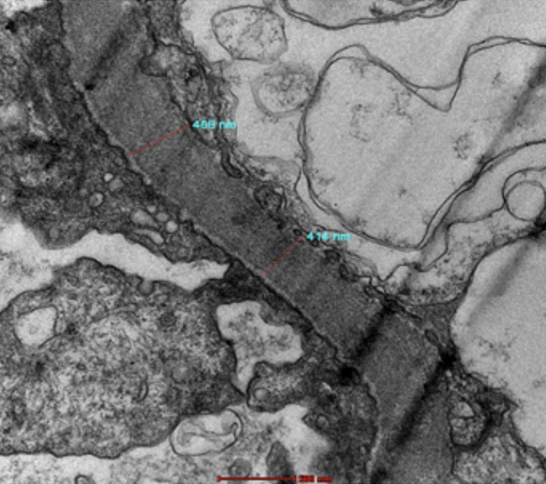
Case report: A 19-year-old male patient with no significant personal medical history but a significant family history of kidney disease: a deceased brother diagnosed with glomerulopathy who refused dialysis, a sister diagnosed with mesangial glomerulopathy, and a mother undergoing dialysis. Genetic testing was declined. He was admitted in May 2022 due to lower-extremity edema and foamy urine persisting for four months. Physical examination revealed hemodynamic stability and bilateral lower-extremity edema. Laboratory results showed: hemoglobin 14.7 g/dL, serum creatinine 0.8 mg/dL, urea 29 mg/dL, serum albumin 2.9 g/dL, and proteinuria 2.03 g/24 hours. Urinalysis showed 15 erythrocytes/µL. Immunological profiles and serology for viral hepatitis, HIV, and VDRL were negative. Serum and urine protein electrophoresis showed no monoclonal component. Evaluation for hematological disorders and computed tomography revealed no significant abnormalities. Targeted genetic testing for hereditary nephrotic syndrome and lysosomal storage disorders yielded negative results. A renal biopsy was performed (Fig. 1 and Fig. 2). Treatment with prednisone was initiated at a dose of 1 mg/kg/day. After two months, given the lack of remission, tacrolimus was added at a dose of 2 mg every 12 hours. Despite treatment, the patient continued to exhibit proteinuria exceeding 7 g/24 hours, with tacrolimus blood levels of approximately 7 ng/mL. At six months, mycophenolate sodium was added at a dose of 360 mg every 12 hours. The clinical course was unfavorable, characterized by persistent nephrotic-range proteinuria and progressive deterioration of renal function. After approximately 18 months of treatment, lab results showed a serum creatinine of 3.7 mg/dL, urea of 141 mg/dL, serum albumin of 3.0 g/dL, creatinine clearance of 33 mL/min, and proteinuria of 7.6 g/24 hours. Finally, on May 16, 2024, he initiated hemodialysis. He was subsequently switched to peritoneal dialysis and is currently on the waiting list for a kidney transplant.
Discussion: Three fundamental characteristics stand out in this case: the early age at diagnosis, the significant familial clustering of renal disease, and the lack of response to multiple immunosuppressive treatments. Although the initial genetic study targeting hereditary nephrotic syndrome was negative, the strong family history necessitates maintaining a high index of suspicion regarding a genetic or familial etiology. A negative result on a genetic panel does not entirely rule out a genetic cause, given the limitations associated with the genes included, unidentified variants, and the state of knowledge at the time the study was performed. Furthermore, resistance to treatment with glucocorticoids, tacrolimus, and mycophenolate raises doubts about a purely immunological etiology mediated by a circulating factor. From a pathological perspective, it is essential to integrate findings from light microscopy, immunofluorescence, and electron microscopy. In this case, the presence of FSGS on light microscopy and diffuse podocytopathy on electron microscopy must be correlated with the clinical presentation and family history to establish the most likely etiological classification.
Conclusions: The rapid progression to end-stage renal disease within approximately two years represents an aggressive clinical course. Prior to kidney transplantation, it is particularly important to determine—to the extent possible—whether the condition is primary FSGS mediated by a circulating factor or a genetic or secondary form, as the risk of recurrence in the renal graft differs considerably among these entities.
Downloads
Published
Issue
Section
License
Copyright (c) 2026 Martin Jesus Gomez Lujan, Alexander Torres Pérez, José Somocurcio Peralta, Roxana Lipa Chancolla. (Author)

This work is licensed under a Creative Commons Attribution-NonCommercial-ShareAlike 4.0 International License.
 10.56867