
¿Está realmente bien posicionado? Tomografía computarizada vs. radiografía de tórax en la evaluación de catéteres de hemodiálisis.
DOI:
https://doi.org/10.56867/Palabras clave:
Posicionamiento de catéter de hemodiálisis, Tomografía, Radiografía de tóraxResumen
Introducción: La posición adecuada de la punta de los catéteres venosos centrales para hemodiálisis es fundamental para garantizar un flujo eficiente y reducir complicaciones como disfunción del acceso, trombosis, arritmias y perforación vascular. la radiografía de tórax es el método más utilizado para la verificación posprocedimiento; sin embargo, la tomografía computarizada (TC) ofrece una mayor precisión anatómica y es considerada el estándar de referencia para la evaluación de la localización de la punta del catéter.
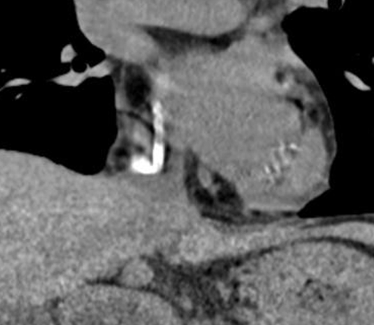
Materiales y métodos: Realizamos un estudio retrospectivo analítico de la base de datos (n 185) de pacientes con diagnóstico de ERC G5 en hemodiálisis, en el periodo enero 2025-junio 2026. Se presentan en 3 grupos: G1: Estenosis centrales (n 116/67%), G2: Agotamiento de los accesos vasculares (n 56/30,2%) y G3: Disfunción del catéter (n 13/7%) . El G3 se sometió a TC para compararla con la Rx convencional de control, realizada de forma rutinaria tras la colocación del dispositivo. Se analizaron las imágenes para determinar la localización de la punta del catéter en conjunto con el especialista en radiología. Se analiza el grupo 3.
Resultados: La edad de los pacientes estuvo entre los 30 y 83 años (media de 62,6) 12 catéteres fueron tunelizados y 1 temporal, 9 ingresaron por vía yugular y 4 por femoral, en 6 se observó que la localización de la punta estaba fuera de la aurícula, 2 se asentaban sobre estenosis de las iliacas primitivas, 1 en la iliaca primitiva izquierda que era cabalgada por la arteria iliaca común (Sn. May Turner) 2 tenían acodadura,1 en el trayecto y otra en la punta del catéter, 1 se localizó en la iliaca externa y en 1 no se encontró la causa de la disfunción. A tres pacientes se les reubicaron los catéteres, retirándoles aproximadamente 3 cm. A 1 no se le realizó ningún procedimiento y a 8 se le colocó un nuevo catéter bajo guía fluoroscópica con control angiográfico.
Conclusión: La tomografía computarizada es más precisa que la radiografía de tórax para determinar la posición de la punta de los catéteres de hemodiálisis y constituye el método de referencia en los estudios de validación. No obstante, su uso debe reservarse para casos complejos o sospecha de malposición.
Descargas
Publicado
Número
Sección
Licencia
Derechos de autor 2026 Favio Arturo Carrera Maigua, Andrea Carolina Buenaño Núñez. (Author)

Esta obra está bajo una licencia internacional Creative Commons Atribución-NoComercial-CompartirIgual 4.0.
 10.56867